Cardiovascular Testing & Diagnostics at Georgia Heart Institute
Heart disease is the leading cause of death for both men and women in the United States, with almost half of all Americans carrying at least one risk factor for heart disease. If you are at elevated risk for heart disease, scheduling a heart scan or diagnostic procedure may save your life. It also ensures that you receive the most precise and in-depth diagnosis possible, which accelerates and improves care.
With a robust team of non-invasive cardiologists and specialty-trained imaging experts, Georgia Heart Institute offers the complete spectrum of heart and vascular diagnostic services through Northeast Georgia Medical Center (NGMC).
Why Choose Georgia Heart Institute?
As Georgia’s most forward-thinking heart program, Georgia Heart Institute uses the most innovative and advanced imaging technology available. This allows us to more thoroughly assess heart disease risk, as well as uncover and treat heart conditions even sooner. Many of our tests are non-invasive, making it easy and convenient to assess your risk and move forward with getting the highly-specialized care you deserve.
All diagnostic services are provided by our expansive and highly-trained team of certified technologists and our board-certified non-invasive cardiologists. Before you receive any diagnostic services, our team will consider a variety of factors to determine the most effective imaging and testing option.
Our Imaging & Diagnostic Services
A variety of different imaging and testing services are available at Georgia Heart Institute through NGMC. Our non-invasive cardiologists and cardiac imaging experts can carefully evaluate the structure of the heart and how it’s functioning, along with the health of the coronary and peripheral arteries. This allows our heart and vascular experts to confirm diagnoses, monitor progress and analyze the effectiveness of treatment.
Nuclear SPECT:
This advanced test works similarly to cardiac PYP by utilizing a radioactive tracer that’s injected into a vein in the arm. That tracer produces gamma rays, which are picked up on by the SPECT camera that rotates around the patient to produce images of the heart. This imaging test is often used to detect blockages in the coronary arteries or to confirm reduced pumping efficiency of the heart.
Cardiac PET:
During this non-invasive test, radioactive tracers capture images of the heart. The images highlight both healthy and unhealthy tissue, allowing cardiologists to diagnose conditions such as coronary artery disease or assess damage after a heart attack.
PET Perfusion (PET/CT):
This test, also called cardiac PET/CT, uses a small amount of radioactivity to capture images of the heart muscle. These images help cardiologists diagnose coronary artery disease while using less radiation than other stress tests.
Cardiac PYP:
This specialized type of testing is used to diagnose a rare condition called cardiac amyloidosis and determine how it can be best treated. During the test, a small amount of a radioactive tracer is injected into a vein in the arm. This tracer produces gamma rays, which are then picked up on images captured by a SPECT/CT camera.
Thallium Viability:
This test is used to evaluate blood flow to the heart following a heart attack or other incident damaging the heart. During this test, a person is safely injected with a radioactive medication called thallium that enables detailed photographs of the heart to be captured over a period of time.
Nuclear Stress Test (Treadmill, Dobutamine & Lexiscan):
- Treadmill Stress Test: During this type of test, a small amount of radioactive tracer is injected, and the patient is connected to specialized equipment that monitors the heart’s function, then asked to begin walking at a slow pace on a treadmill. Over time, the pace and incline are increased, making the heart work harder. The test measures how efficiently the heart is working when stressed or under exertion, as well as at rest.
- Dobutamine Stress Test: When a patient is unable to safely exercise, this type of stress test may be used. Instead of walking or running on a treadmill to stress the heart, this procedure involves injecting a medication called dobutamine, which causes the heart to beat faster, mimicking how the body works during physical activity.
- Lexiscan Stress Test: This test works similarly to a dobutamine stress test, except a medication called lexiscan is injected to stimulate the heart.
Cardiac
Stress Test (Treadmill & Dobutamine)
- Treadmill Stress Echo: During stress echocardiography, a person exercises on a treadmill or bike while his or her blood pressure and heart rhythm are monitored. When the person’s heart rate reaches a peak level, a doctor captures ultrasound images of the heart to determine how well the heart and blood vessels are working.
- Dobutamine Stress Echo: This test works similarly to a standard stress echo, but uses a medication called dobutamine to stimulate the heart instead of exercise.
Transthoracic Echo (TTE):
During a transthoracic echocardiogram, a technician uses an instrument called a transducer to capture images of the heart through the chest or abdominal wall. The transducer sends sound waves into the chest, then picks up the echoes that reflect off different parts of the heart.
Transesophageal Echo (TEE):
Transesophageal echocardiogram works similarly to a transthoracic echo, except the transducer is placed in the esophagus. This provides clear images of the heart and its structures.
Vascular
Aorta IVC Iliac Complete:
This test uses ultrasound to take a detailed look at the aorta and other arteries that deliver blood throughout the body. This includes an evaluation of the iliac arteries, which supply oxygenated blood to the pelvis, organs located in the pelvis, and the reproductive organs.
Mesenteric Artery Study:
This test uses ultrasound to check the function of the mesenteric arteries, which branch off the abdominal aorta. It is used to capture images that help identify stenosis, plaque buildup, or occlusions that can disrupt blood flow to the intestines, the liver, the stomach, or the spleen.
Arterial Duplex Scan:
This test uses ultrasound to capture detailed images of the major arteries in the arms, legs, and neck. It is typically used to identify plaque buildup and blockages that may lead to conditions such as peripheral arterial disease, or PAD.
Venous Duplex Scan:
This test uses ultrasound to capture images of the veins, allowing a doctor to evaluate how efficiently blood is flowing through the venous system.
Carotid Duplex Scan:
This test uses ultrasound to capture images of the carotid arteries, the arteries that run from the heart up through the neck and into the brain.
Hemodialysis Access Duplex Scan:
This specialized type of scan is performed on a person who is undergoing dialysis. The test captures images of the dialysis access site, checking for arterial outflow, conduit, and central venous outflow, helping to ensure optimal placement and output for dialysis.
Portal Vein Duplex Scan:
This test uses ultrasound to capture images of the hepatic portal vein, which moves blood from the spleen and gastrointestinal tract to the liver.
Transcranial doppler:
This test uses ultrasound to capture images that help detect problems with the blood flow in the brain. It can be used to identify stroke, narrowed blood vessels, hemorrhage, and blood clots.
Vein Mapping:
This test uses ultrasound to capture images of the veins, allowing a doctor to determine whether there are any blockages. Vein mapping is often used in advance of certain medical procedures, such as open heart surgery, to determine whether there are veins sufficient enough to be used as grafts.
Ankle Brachial Index (ABI) Screening:
This test uses ultrasound in conjunction with blood pressure monitoring to determine whether there’s a potential narrowing or blocking of the arteries in the legs. During the test, blood pressure is checked in both the arms and legs and compared to determine the quality of blood flow in the ankles.
Pulse Volume Recording (with Treadmill Exercise):
Similar to ankle brachial index screening, this test uses ultrasound to examine blood flow in the arms and legs. In some cases, images may be captured while a person is at rest and while walking or running on a treadmill.
Holter Monitor (24-hour and 48-hour):
This type of test is essentially a portable electrocardiogram. The device is worn for either a 24-hour or 48-hour interval, and during that time, it records the electrical activity of the heart. Holter monitoring is often recommended to help make or confirm a diagnosis of a heart health issue such as an arrythmia.
Cardiac Event Monitor:
This test works similarly to Holter monitoring, but has two main differences. It does not record electrical activity continuously; instead, it is often set to record when an abnormal heart rhythm is detected. This type of monitor is also worn for longer periods of time.
Cardiac Stress:
During this test, MRI captures detailed images of the heart while it’s both at rest and while stressed. A person is injected with medication to mimic the way blood flows in the heart during exercise. Contrast dye is then injected into a vein, helping to identify any areas of the heart without proper blood flow.
Cardiac MRI:
This test also uses MRI and sometimes contrast dye to get a clear look at the heart and blood vessels around the heart, but unlike a stress MRI, this test looks only at the heart while it’s at rest.
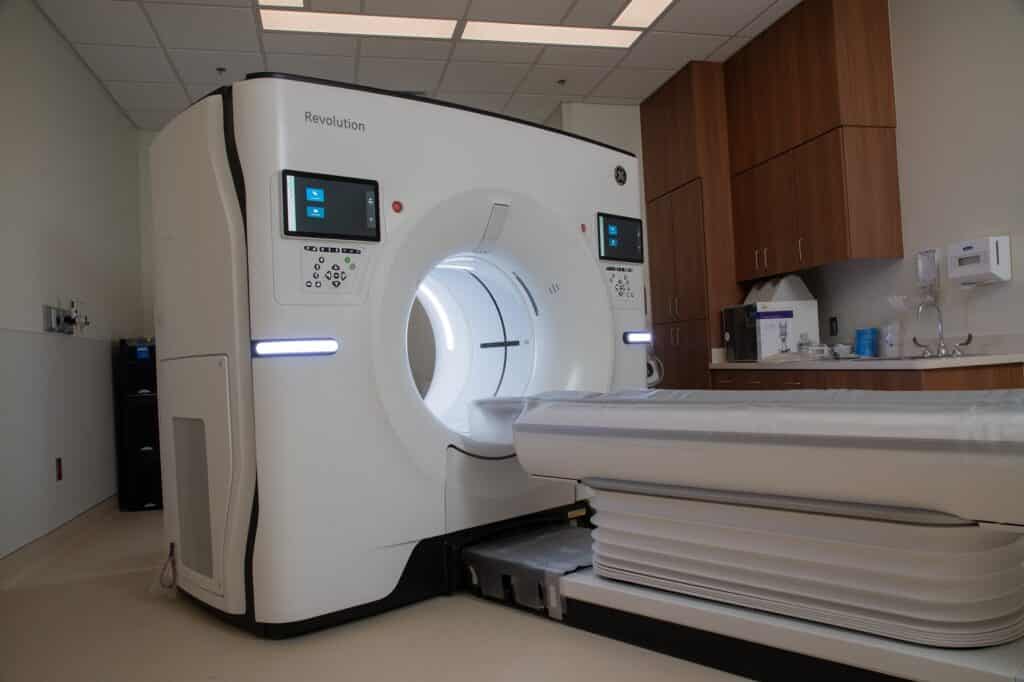
Cardiac CT Angiogram:
This test, sometimes called cardiac CT angiography, is used to identify whether the arteries are blocked by plaque buildup. During the CCTA, a person is injected with contrast dye and then computed tomography (CT) is used to capture 3D images of the arteries supplying blood to the heart.
Coronary Calcium Scoring:
This test, which is also called a “heart scan,” uses a CT scan to measure the amount of calcified plaque in the coronary arteries. The calcium score is used to predict the likelihood of a coronary event.
HeartFlow Analysis (CT FFR):
This test uses computed tomography to create a color-coded 3D model of the coronary arteries. This model helps identify where and how blockages are impacting blood flow.
Arterial CT Scan (IVC, Aorta & Iliac):
This test uses computed tomography to capture images of the arteries and veins, including the inferior vena cava, the aorta, and the iliac arteries, which supply oxygenated blood to the pelvis, organs located in the pelvis, and the reproductive organs.
FAQs
Because Georgia Heart Institute and Northeast Georgia Medical Center offer an extensive array of heart and vascular imaging services, your cardiologist and/or advanced practice provider will coordinate all scheduling for you. This ensures that you receive the best imaging tests for your heart and vascular needs. Once an imaging order has been submitted by your care expert, you can contact one of our locations if you have any questions or need to schedule your service.
Each type of imaging uses different technology or methods to produce images of the heart and the surrounding blood vessels. Several types of imaging tests involve a low level of radiation exposure. This is minimized by utilizing the most up-to-date technology and processes. All of our imaging technicians, radiologists and non-invasive cardiologists are specially trained to ensure your health and safety while undergoing any imaging procedure.
Locations
Schedule an Appointment
Start your journey toward lasting heart health today! Simply schedule your appointment with one of our experts online or call the location closest to you!